- How Blue Light Impacts Your Sleep Quality
- Comparing Whey and Plant-Based Protein: Which is Best?
- How Long Does Nicotine Remain in Your System?
- The Best Time of Day to Drink Bone Broth to Maximize Health Benefits
- 8 Ways to Increase Dopamine Naturally
- 7 Best Breads for Maintaining Stable Blood Sugar
- Gelatin vs. Collagen: Which is Best for Skin, Nails, and Joints?
- The Long-Term Effects of Daily Turmeric Supplements on Liver Health
- Could Your Grocery Store Meat Be Causing Recurring UTIs?
- Are You Making This Expensive Thermostat Error This Winter?
Treatments for Brain Cancer Take Heavy Toll on the Brain
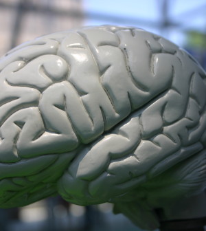

Radiation and chemotherapy can cause structural changes in the healthy brain tissue of patients with glioblastoma brain tumors, a new study finds.
The research included 14 glioblastoma patients who underwent 35 weeks of combined radiation and chemotherapy (chemoradiation) after having their tumors surgically removed.
The patients had brain scans before and after chemoradiation, but an adequate number of images were obtained from only eight of the patients. Those images revealed a significant decrease in whole brain volume — the overall amount of brain tissue — throughout chemoradiation.
The reduced amount of brain tissue became apparent within a few weeks after the start of chemoradiation and was primarily seen in gray matter.
The scans also showed that the size of the brain’s ventricles — cerebrospinal fluid-filled spaces deep within the brain — grew progressively larger during chemoradiation. Changes were also detected within the subventricular zone, one of two structures in which new brain cells are generated in adults.
The study was published recently in the journal Neurology.
It was well known that whole brain radiation can have harmful effects on the brain. But, this is the first study to look at structural brain changes caused by chemoradiation, according to senior study author Dr. Jorg Dietrich, of the Pappas Center for Neuro-Oncology at Massachusetts General Hospital in Boston.
“We were surprised to see that these changes — reduced gray matter volume and ventricular enlargement — occurred after just a few weeks of treatment and continued to progress even after radiation therapy was completed,” he said in a hospital news release.
“While this was a small study, these changes affected every patient at least to some degree. Now we need to investigate whether these structural changes correlate with reduced cognitive function, and whether neuroprotective strategies might be able to stop the progression of brain volume loss,” said Dietrich, an assistant professor of neurology at Harvard Medical School.
More information
The U.S. National Cancer Institute has more about brain tumors.
Source: HealthDay
Copyright © 2026 HealthDay. All rights reserved.
Related
-
 Navigating Your Midlife Crisis: Embracing New Possibilities
A midlife crisis can open doors to personal growth...
- December 4, 2025
-
 Want A Happier Hospital? Hire More Nurses, Study Says
Adding even a few extra nurses can dramatically reduce...
- November 20, 2025
-
 Plasma Treatment Shows Promise For Menopause Symptoms Among Breast Cancer Survivors
Plasma infusions may help breast cancer survivors avoid genital...
- November 20, 2025
-
 Self-Hypnosis Can Thwart Hot Flashes
Self-hypnosis might help some women in menopause find relief...
- November 14, 2025


