- 8 Ways to Increase Dopamine Naturally
- 7 Best Breads for Maintaining Stable Blood Sugar
- Gelatin vs. Collagen: Which is Best for Skin, Nails, and Joints?
- The Long-Term Effects of Daily Turmeric Supplements on Liver Health
- Could Your Grocery Store Meat Be Causing Recurring UTIs?
- Are You Making This Expensive Thermostat Error This Winter?
- Recognizing the Signs of Hypothyroidism
- 10 Strategies to Overcome Insomnia
- Could Artificial Sweeteners Be Aging the Brain Faster?
- Techniques for Soothing Your Nervous System
Preemies Show Subtle Differences in Brain Development


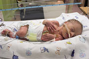
Premature infants with no obvious problems in the structure of their brains may still have subtle chemical differences compared with full-term babies, a new study finds.
Researchers said it’s not clear if these microscopic differences are actually signs of trouble. But they hope that a deeper understanding of preemies’ brain development will eventually be useful in improving their outlook.
“Many premature infants are healthy, but they are at increased risk of problems,” said lead researcher Stefan Bluml, of Children’s Hospital Los Angeles. Those problems can include learning disabilities, behavioral issues — such as attention-deficit/hyperactivity disorder — and autism.
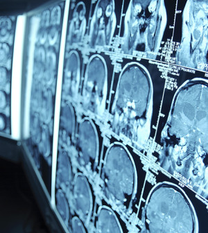
To gain more insight into preemie brain development, Bluml’s team used magnetic resonance spectroscopy (MRS) to image infants’ brains on the microscopic level.
All 81 babies in the study — 30 preterm and 51 full-term — had normal brain structure, based on standard MRI scans. But when the researchers used MRS, they saw “biochemical” differences between the premature and full-term infants.
In general, the preemies showed an “early start” in the brain’s white matter development, which put it out of sync with the maturation of the brain’s gray matter. Gray matter can be seen as the brain’s information-processing centers, while white matter is like the wiring connecting those centers.
Bluml described it as a “false start” in preemies’ white matter development, and the trigger appears to take place after birth. It’s not clear what that trigger is, but one possibility, according to Bluml, is oxygen.
The fetal brain, he explained, is designed to develop in a low-oxygen environment. At birth, babies are thrust into a much more oxygen-rich world, and preemies’ brains might not be quite ready for that.
Still, it’s not clear if the brain differences Bluml’s team found are “bad.” “These might be necessary compensatory mechanisms,” Bluml said. “At present, it’s not clear. We’re just saying there are differences.”
The findings are scheduled for presentation Sunday at the Radiological Society of North America annual meeting, in Chicago. Research presented at meetings should be viewed as preliminary until published in a peer-reviewed medical journal.
One expert agreed that the significance is unknown. But it would be interesting to follow these infants over time, to see if the chemical differences are associated with any learning or behavior issues later, said Dr. Madhavi Koneru, a neonatologist who treats newborns at McLane Children’s Hospital at Scott & White in Temple, Texas.
“What does this mean down the road? That’s what we want to know,” Koneru said.
In recent years, medical advances have allowed more and more of the tiniest preemies to survive. But, Koneru said, “surviving is not enough. We want them to thrive.”
She said she thinks that brain-imaging research like this will one day aid in improving preemies’ outcomes.
For now, Bluml said, they are just trying to map the course that preemies’ brain maturation typically takes. “Our goal is to establish certain landmarks of brain development,” he said.
Eventually, he added, MRS scans could potentially be used to evaluate whether a therapy for preterm babies is actually having an effect on brain development.
More information
The March of Dimes has more on preterm birth.
Source: HealthDay
Copyright © 2026 HealthDay. All rights reserved.


