- How Blue Light Impacts Your Sleep Quality
- Comparing Whey and Plant-Based Protein: Which is Best?
- How Long Does Nicotine Remain in Your System?
- The Best Time of Day to Drink Bone Broth to Maximize Health Benefits
- 8 Ways to Increase Dopamine Naturally
- 7 Best Breads for Maintaining Stable Blood Sugar
- Gelatin vs. Collagen: Which is Best for Skin, Nails, and Joints?
- The Long-Term Effects of Daily Turmeric Supplements on Liver Health
- Could Your Grocery Store Meat Be Causing Recurring UTIs?
- Are You Making This Expensive Thermostat Error This Winter?
Alzheimer’s May Vary, Brain to Brain
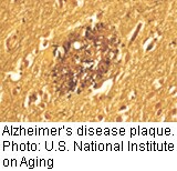
THURSDAY, Sept. 12Differences in plaque-forming structures in the brains of Alzheimer’s patients may offer clues to why the disease can progress more rapidly or be less severe in some people, a new study suggests.
The research could spur the development of new imaging agents that highlight specific structures in the brain — called beta-amyloid fibrils — improving the reliability and specificity of diagnosis, according to Robert Tycko, lead author of the paper published Sept. 12 in the journal Cell.
“Variations in disease may have a structural basis and be due to differences in the molecular structure of the fibrils,” said Tycko, a senior investigator with the intramural research program of the U.S. National Institutes of Health.
The study represents an important advance in Alzheimer’s research, an expert not involved with the study noted.
Beta-amyloid fibrils are responsible for the amyloid plaques seen in Alzheimer’s disease, the most common form of dementia. Alzheimer’s is the sixth leading cause of death in the United States and more than 5 million Americans have the disease, according to the Alzheimer’s Association.
For the research, the scientists took tissue from the brains of two deceased female Alzheimer’s patients with different signs and symptoms of the disease. They extracted beta-amyloid from the tissue and used it as “seeds” to grow beta-amyloid fibrils. The investigators found that the same “seeds” — the amino acid sequence — could assemble into different molecular structures.
Using nuclear magnetic resonance and electron microscopy to visualize the beta-amyloid fibrils in the patients’ brain tissue, the scientists discovered correlations between variations in the disease and differences in molecular structure.
“There are at least two different varieties [of amyloid structure] in Alzheimer’s disease,” Tycko said. “And certain fibril structures may be more likely than others to cause the disease.”
Tycko explained that while the research team was able to determine that there are at least two structural varieties of Alzheimer’s disease, they were unable to prove that there are correlations between variations in disease and molecular structure.
He said he hopes that the research will eventually lead to the ability to tell someone with memory loss whether or not the problem is likely to lead to a serious or fast-moving form of Alzheimer’s.
One expert not involved with the research called the discovery a “technical tour de force.”
“The research is a huge step forward,” said Terrence Town, a professor of physiology and biophysics at the Keck School of Medicine at the University of Southern California. “They have accomplished something we have been trying to do for a decade.”
For years, researchers have been focusing on something smaller than a fibril, called an oligomer, considered to be especially toxic to the brain, Town explained. “Now this paper is drawing attention to something different: fibrils.”
The findings will help researchers focus on the fibrils, ideally working toward developing ways to identify and diagnose people in the earliest stages of the disease, Town said.
More information
Learn more about Alzheimer’s disease from the U.S. Centers for Disease Control and Prevention.
Source: HealthDay
Copyright © 2026 HealthDay. All rights reserved.
Related
-
 Navigating Your Midlife Crisis: Embracing New Possibilities
A midlife crisis can open doors to personal growth...
- December 4, 2025
-
 Want A Happier Hospital? Hire More Nurses, Study Says
Adding even a few extra nurses can dramatically reduce...
- November 20, 2025
-
 Plasma Treatment Shows Promise For Menopause Symptoms Among Breast Cancer Survivors
Plasma infusions may help breast cancer survivors avoid genital...
- November 20, 2025
-
 Self-Hypnosis Can Thwart Hot Flashes
Self-hypnosis might help some women in menopause find relief...
- November 14, 2025


