- FDA Says First Round of Tests Show No Live Virus in Pasteurized Milk
- King Charles Returns to Duties After Cancer Treatment
- Biden Administration Delays Menthol Cigarette Ban
- Blood Test Might Predict Knee Osteoarthritis Years Early
- Dogs Can Get Lyme Disease, Too
- Spinal Cord Injury Damages Metabolism, and Scientists Now Know Why
- Syphilis Is Increasingly Displaying Atypical, Severe Symptoms
- Climate Change Could Be Good News for Viruses Like COVID
- Vaccines Have Saved 154 Million Lives, Mostly Babies, Over Past 50 Years
- Scientists Discover Cause of Rare Movement Disorder
Mammograms: An Expert Overview on Why They’re So Important

Mammograms have long offered early detection of breast cancer, which is why getting them regularly is crucial to women’s health, one expert says.
“There are several risk factors associated with breast cancer. As with many other diseases, risk of developing breast cancer increases as you get older,” said Dr. Mridula George, associate program director of breast medical oncology at Rutgers Cancer Institute of New Jersey.
Breast cancer is the second-most common cancer for women after skin cancer, according to the American Cancer Society.
A woman whose mother or sister developed breast or ovarian cancer may be at high risk for the disease. So, too, might someone who has multiple family members who developed breast, ovarian or prostate cancer.
In the early stages of breast cancer, it may not be possible to find signs through breast self-exam. Early disease also doesn’t cause pain, George noted in a Rutgers news release.
Later, symptoms can include a lump or thickening in or near the breast or in the underarm area. It may be seen as a change in the size or shape of the breast or felt as tenderness.
A woman may also experience nipple discharge or the nipple pulled back into the breast, or a change in the way the skin of the breast, areola or nipple looks or feels, such as being warm, swollen, red or scaly, George added.
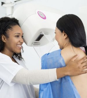
Mammography uses low-dose X-rays to show abnormal areas or tissues in the breast before a woman has noticeable symptoms.
The breasts are each placed in a special machine between two plates. The plates move together to compress the breast tissue, so it’s easier for the X-ray to obtain a clear image.
The images are stored on a computer where they can be viewed and analyzed by the radiologist and a woman’s doctor.
When a breast cancer is detected early and hasn’t spread, the five-year relative survival rate is 99%, George said. Those found during screening exams are more likely to be smaller and less likely to have spread outside the breast.
A woman can talk to her doctor about when to start screenings.
More information
The U.S. National Cancer Institute has more on mammograms.
SOURCE: Rutgers Cancer Institute of New Jersey, news release, Oct. 1, 2023
Source: HealthDay
Copyright © 2024 HealthDay. All rights reserved.


