- How Blue Light Impacts Your Sleep Quality
- Comparing Whey and Plant-Based Protein: Which is Best?
- How Long Does Nicotine Remain in Your System?
- The Best Time of Day to Drink Bone Broth to Maximize Health Benefits
- 8 Ways to Increase Dopamine Naturally
- 7 Best Breads for Maintaining Stable Blood Sugar
- Gelatin vs. Collagen: Which is Best for Skin, Nails, and Joints?
- The Long-Term Effects of Daily Turmeric Supplements on Liver Health
- Could Your Grocery Store Meat Be Causing Recurring UTIs?
- Are You Making This Expensive Thermostat Error This Winter?
3-D Model of Heart May Help Surgeons Fix Defects

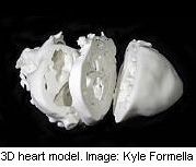
Being able to examine a 3-D model of the heart may boost surgeons’ ability to treat patients born with complex cardiac defects, a new study suggests.
Heart surgeons typically rely on 2-D images taken by X-ray, ultrasound or MRI to plan their surgery on a patient. But these images may not reveal complex structural defects in the heart present at birth, the researchers explained.
But now, advances in technology are enabling surgeons to build and print detailed 3-D models of patients’ hearts from plaster, ceramic or other materials in order to gain a full understanding of what they’ll face during surgery.
Researchers used the new technology in treating three patients who were born with complex heart defects. In each case, the 3-D model provided important information that wasn’t available from traditional imaging and that influenced how the surgery was performed.
The heart abnormalities were repaired in all three patients, according to the study, which was to be presented on Wednesday at the annual meeting of the American Heart Association (AHA) in Chicago.
“With 3-D printing, surgeons can make better decisions before they go into the operating room,” lead author Dr. Matthew Bramlet, director of the Congenital Heart Disease MRI Program at the University of Illinois College of Medicine, said in an AHA news release.
“The more prepared they are, the better decisions they make, and the fewer surprises that they encounter,” he added.
“When you’re holding the heart model in your hands, it provides a new dimension of understanding that cannot be attained by 2-D or even 3-D images,” he said.
The researchers stressed that this approach is still new and that 3-D printing has not been approved by the U.S. Food and Drug Administration. Findings presented at medical meetings are also typically considered preliminary until published in a peer-reviewed journal.
More information
The U.S. National Heart, Lung, and Blood Institute has more on congenital heart defects.
Source: HealthDay
Copyright © 2026 HealthDay. All rights reserved.
Related
-
 Navigating Your Midlife Crisis: Embracing New Possibilities
A midlife crisis can open doors to personal growth...
- December 4, 2025
-
 Want A Happier Hospital? Hire More Nurses, Study Says
Adding even a few extra nurses can dramatically reduce...
- November 20, 2025
-
 Plasma Treatment Shows Promise For Menopause Symptoms Among Breast Cancer Survivors
Plasma infusions may help breast cancer survivors avoid genital...
- November 20, 2025
-
 Self-Hypnosis Can Thwart Hot Flashes
Self-hypnosis might help some women in menopause find relief...
- November 14, 2025


